INC-IEM Neuroengineering Seminar Series
Organized by:
Institute for Neural Computation and Institute of Engineering in Medicine
Coming Up
TBA
When: , 2026,
2:00pm - 3:00pm
Where: Hybrid
In-person: Fung Auditorium, Powell-Focht Bioengineering Hall Rm 191
Over Zoom: https://ucsd.zoom.us/j/2888083696
Meeting ID: 288 808 3696
Past Talks
2026-05-27
Neuroadaptive AI: Using Brain-Derived Signals to Ground LLMs and Physical AI in Human Cognitive State
Thorsten O. Zander Lichtenberg Chair for Neuroadaptive HCI, BTU, Germany - https://www.b-tu.de/en/fg-neuroadaptive-hci/page
CLICK HERE to view the recorded talk
Abstract: Neuroadaptive technology uses brain activity as an additional source of implicit information about how a user perceives, evaluates, and understands an ongoing interaction (Zander et al., 2016). Rather than treating brain signals as explicit commands, passive BCI systems can estimate covert user states such as attention, workload, error processing, surprise, or agreement, and use these states to adapt interactive technology (Zander & Kothe, 2011). In this talk, I will present recent work on neuroadaptive AI, with a focus on progress made in the NAFAS project. NAFAS aims to develop robust, wearable, and scalable passive BCI technology for real-world interaction contexts, including advances in usable EEG sensing, transferable classifiers, and continuous user-state estimation. I will then discuss how such signals can be used as structured annotation traces for LLM-based and physically embodied AI systems. The central idea is that brain-derived signals can augment the information space available to AI systems, helping them infer user beliefs, detect misalignment, estimate uncertainty, and adapt interaction strategies. I will argue that this provides a pragmatic path toward more context-aware and human-compatible AI, while also raising important scientific and ethical challenges related to robustness, interpretability, privacy, and evaluation.
Biography: Prof. Dr. Thorsten O. Zander is a Lichtenberg Professor for Neuroadaptive Human-Computer Interaction at Brandenburg University of Technology Cottbus-Senftenberg, Germany and Co-Founder of Zander Labs, Berlin, Germany. He introduced and shaped the concept of passive brain-computer interfaces and neuroadaptivity, where brain activity is used to infer covert user states and adapt interactive systems without explicit control commands. His work spans neuroadaptive technology, passive BCI, cognitive state monitoring, human-machine interaction, and brain-derived information channels for human-compatible AI systems.
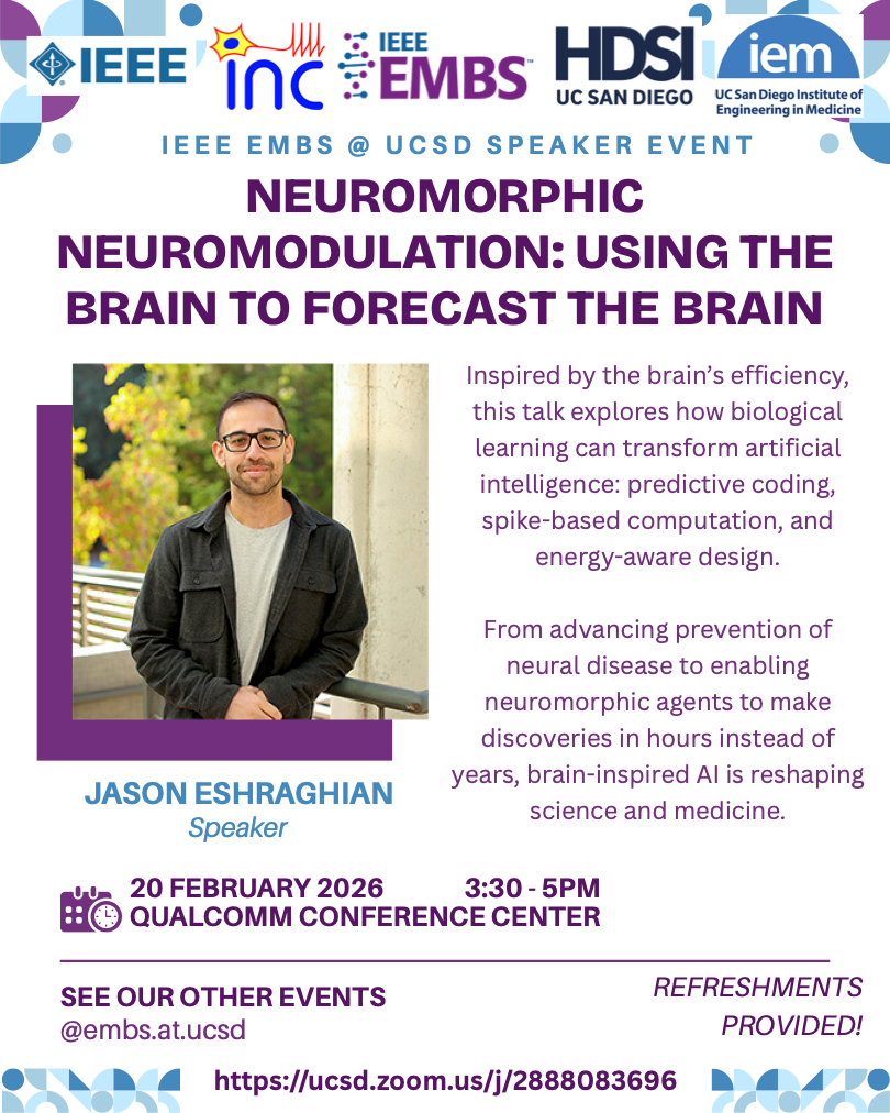
2026-02-20
Neuromorphic Neuromodulation: Using the Brain to Forecast the Brain
Jason Eshraghian University of California, Santa Cruz - Event Flyer
CLICK HERE to view the recorded talk
Abstract: The brain is nature's most efficient computer, and the perfect blueprint for building better neural networks. This talk explores how principles of biological learning can revolutionize artificial intelligence: predictive coding that anticipates rather than reacts, spike-based computation that processes information through discrete events, and energy-aware learning that mirrors how the brain optimizes for minimal metabolic cost.
We apply these principles to neuromorphic neuromodulation, enhancing our ability to forecast and take preventative action against neural disease. But the implications extend beyond medicine: by translating these biological insights into computational frameworks, we enabled neuromorphic agents to make scientific discoveries in 2.5 hours that took the neuromorphic computing community half a decade to uncover.
Biography: Jason Eshraghian is an Assistant Professor and Fulbright Scholar in the Department of Electrical and Computer Engineering at the University of California, Santa Cruz. He is the developer of snnTorch, a Python library with over 500,000 downloads for training spiking neural networks. He is a dual-appointed IEEE CAS and EMBS Distinguished Lecturer, an Associate Editor of APL Machine Learning, the Chair of the IEEE Neural Systems and Applications Technical Committee, has been the recipient of seven IEEE Best Paper Awards, a Scientific Advisory Board Member of BrainChip and leads the Neuromorphic Agents Team at Conscium.
2025-09-24
Mapping the Rules of Neurological Disorders Using Single Cell and Spatial Genomics
Omer Ali Bayraktar Wellcome Sanger Institute, Hinxton, United Kingdom - https://www.sanger.ac.uk/group/bayraktar-group/
CLICK HERE to view the recorded talk
Abstract: Single cell and spatial transcriptomics promise to resolve the cellular wiring diagrams of tissues in health and disease, but it remains challenging to extract biological insight from complex human tissue atlases. Here, I will demonstrate how we use multi-modal tissue atlassing to discover the rules of two neurological disorders. First, I will talk about our spatial transcriptomic atlas of genetic risk genes for autism spectrum disorder (ASD) in the developing human brain. Leveraging single cell transcriptomics, we identified region ASD gene programs and discovered convergence of ASD risk genes in thalamic circuits. Second, I will present GBM-space, a multi-modal map of glioblastoma tumour tissue architecture. By deeply profiling tumours across multiple sites using integrated single cell and spatial genomics, we identified a novel cellular and tissue framework unifying glioblastoma tumour heterogeneity. These efforts show how cell atlassing can discover the fundamental principles of complex diseases.
Biography: Dr. Bayraktar uses single cell and spatial transcriptomics technologies to characterize human brain cellular diversity in health and disease. During his training, Omer discovered neural stem cell patterning mechanisms (Nature 2013) and astrocyte layer diversity in the cerebral cortex (Nature Neuro 2020). Omer started his independent research group at the Wellcome Sanger Institute in 2018. His team has developed the cell2location computational tool to map fine cell types in spatial transcriptomics (Nature Biotech 2022). Omer is internationally funded by Wellcome LEAP, SFARI and CZI, and he steers spatial genomic strategy at the Sanger Institute.
2025-07-23
Miniaturized Neural Engineering Systems Enabled by Energy-Efficient Integrated Circuits
Chul Kim Korea Advanced Institute of Science & Technology (KAIST) - https://beee.kaist.ac.kr/
CLICK HERE to view the recorded talk
Abstract: As the global population continues to age and humanity faces an increasing number of previously unencountered diseases, the importance of neural engineering has grown significantly. Among the various approaches within neural engineering, miniaturized systems based on semiconductor integrated circuit technology are emerging as a critical component. This presentation introduces a series of research efforts aimed at addressing biomedical challenges through highly energy-efficient integrated circuit systems. Specifically, we will highlight innovations in low-power neural recording circuits, real-time removal of electrical stimulation artifacts, wireless power and data transmission systems that are robust to distance variations, and unobtrusive wearable devices. These advancements demonstrate the transformative potential of semiconductor technologies in the future of biomedical applications.
Biography: Chul Kim (Senior Member, IEEE) received the Ph.D. degree in Bioengineering from UC San Diego, La Jolla, CA, USA, in 2017. He is an Associate Professor with the Department of Bio and Brain Engineering and the Program of Brain and Cognitive Engineering, Korea Advanced Institute of Science and Technology (KAIST), Daejeon, South Korea. From 2009 to 2012, His research interests include the design of energy-efficient integrated circuits and systems for next-generation brain-machine interfaces, electroceuticals, and unobtrusive wearable sensors. He received the Gold Prize in the 16th Humantech Thesis Prize Contest from Samsung Electronics, Suwon, South Korea, in 2010, and the 2018 Shunichi Usami Ph.D. Thesis Design Award from the Bioengineering Department at UC San Diego. He served as an Associate Editor of IEEE Transactions on Biomedical Circuits and Systems (TBioCAS) and on the Technical Program Committee for IEEE Custom Integrated Circuits Conference (CICC) as Co-Chair of the Emerging Technology Sub-committee.
2025-05-02
Who Will Design Tomorrow’s Analog Integrated Circuits: Humans Or AI-based Synthesis?
Georges Gielen KU Leuven, Belgium
CLICK HERE to view the slides
Abstract: Analog/mixed-signal integrated circuits are key in applications where electronics interface with the physical world. The design of analog circuits, however, is time consuming and prone to errors, often requiring multiple redesign cycles. The rebirth of AI and machine learning, and the recent rise of generative AI methods, on the other hand, create a whole new spectrum of techniques to automate this process. This invited talk will explore the high potential of using advanced machine learning (ML) techniques to automatically synthesize and lay out analog integrated circuits. What is hype and what will be feasible? Will we still need analog designers in the future and how will they operate?
Biography: Georges G.E. Gielen received the MSc and PhD degrees in Electrical Engineering from the Katholieke Universiteit Leuven (KU Leuven), Belgium, in 1986 and 1990, respectively. Currently, he is Full Professor in the MICAS research division at the Department of Electrical Engineering (ESAT) at KU Leuven. From 2013 until 2017 he served as Vice-Rector for the Group of Sciences, Engineering and Technology. In 2018 he was visiting professor at UC Berkeley and Stanford University. From 2020 to 2024 he served as Chair of the Department of Electrical Engineering (ESAT) at KU Leuven. His research interests are in the design of analog and mixed-signal integrated circuits, and especially in analog and mixed-signal CAD tools and design automation, including modeling, simulation, optimization and synthesis as well as testing. He has graduated over 55 PhDs so far. He is a frequently invited speaker and serves/served as Distinguished Lecturer of the IEEE Solid-State Circuits Society and the IEEE Circuits and Systems Society. He is coordinator/partner of several academic and industrial research projects in the above fields, including having awarded the ERC Advanced Grant AnalogCreate. He has (co-)authored 14 monograph books and more than 800 publications in edited books, international journals and conference proceedings. He is a 1997 Laureate of the Belgian Royal Academy of Sciences, Literature and Arts in the discipline of Engineering. He is Fellow of the IEEE since 2002, and received the IEEE CAS Mac Van Valkenburg award in 2015, the IEEE CAS Charles Desoer award in 2020, as well as the EDAA Achievement Award in 2021. He is an elected member of the Royal Flemish Academy of Belgium in the class of Technical Sciences, and of the Academia Europaea.
2025-01-15
Regulation without Calibration
Rodolphe Sepulchre KU Leuven, Belgium, and University of Cambridge, UK, https://sites.google.com/site/rsepulchre/
CLICK HERE to view the recorded talk
Abstract: Regulation theory is grounded in the internal model principle, which states that exact regulation requires an exact internal model of the external signals to be regulated. How to reconcile this calibration principle with systems made of uncertain and variable components? How do animals achieve regulation in changing and complex environments? The talk will propose that reliable regulation is possible in uncertain machines that regulate events rather than trajectories. I will highlight the role of excitability and synaptic coupling in a theory of event regulation.
Biography: Rodolphe Sepulchre is Professor of Engineering at the KU Leuven (Belgium) and at the University of Cambridge (UK). He is a fellow of IFAC (2020), IEEE (2009), and SIAM (2015). He received the IEEE CSS Antonio Ruberti Young Researcher Prize in 2008 and the IEEE CSS George S. Axelby Outstanding Paper Award in 2020. He was elected at the Royal Academy of Belgium in 2013. He has been Editor-in-Chief of Systems and Control Lettters (2009-2019) and the IEEE Control Systems Magazine (2020-2024). He is a recipient of two ERC advanced grants (Switchlets (2015-2021) and SpikyControl (2023-2028).
2024-11-18
Distributed and Localized Model Predictive Control
Carmen Amo Stanford University https://camoalon.github.io
CLICK HERE to view the recorded talk
Abstract: The increasing presence of large-scale distributed systems highlights the need for scalable control strategies where only local communication is required. Moreover, in safety-critical systems it is imperative that such control strategies handle constraints in the presence of disturbances and enjoy theoretical and performance guarantees. In response to this need, we present the Distributed and Localized Model Predictive Control (DLMPC) algorithm for large-scale linear systems. DLMPC is a distributed closed-loop model predictive control (MPC) scheme wherein only local state and model information needs to be exchanged between subsystems for the computation and implementation of control actions. The resulting distributed algorithms tackle various types of additive disturbances and enjoy recursive feasibility and asymptotic stability guarantees that introduce minimal conservatism and can be computed in an offline fashion without adding to the computational burden. We also provide analysis and guarantees on the global performance of DLMPC, and demonstrate that in cases where the underlying topology of the system is sparse (as is the case in most large-scale networks), the inclusion of local communication constraints does not result in a suboptimal solution. Moreover, we show that when no noise is present, this algorithm can be extended to the purely data-driven case where all previous guarantees hold and the need for a model is fully replaced by past-trajectory data. We show that the amount of data needed for our synthesis problem is independent of the size of the global system. Lastly, we explore the potential of DLMPC for hardware accelerated implementation in GPU by exploiting the fact that the structure of the DLMPC problem captures some of the limitations of GPU computations. In all algorithmic and theoretical results presented in this talk, only local information exchange is necessary, and computational complexity is independent of the global system size. DLMPC is the first MPC algorithm that allows for the scalable, efficient and data-driven computation and implementation of distributed closed-loop control policies and enjoys theoretical guarantees.
2024-04-16
Bio/Nano/CMOS Interfaces for Remote Monitoring of Human Metabolism
Sandro Carrara École Polytechnique Fédérale de Lausanne (EPFL), Switzerland, https://www.epfl.ch/labs/bci/
CLICK HERE to view the recorded talk
Abstract: IoT wearable-devices (accelerometers, heartbeat monitoring systems, etc) are key for better, more rational, effective and ultimately low-cost health care at home. However, these systems do not usually allow measurement of metabolites at the molecular level. So far, there are no available integrated nano-bio-systems for multi-metabolites, real-time, remote monitoring of human metabolism. This talk presents innovative concepts for multi-panel, highly integrated, fully implantable, remotely powered and real-time molecular-level monitoring of human metabolism. Applications are shown in the field of implantable and wearable devices, with in-vivo experiments, and addressing biocompatible-packaging, flexible electronics, and monitoring-needs in intensive care units. Future perspective on injectable body dust devices are addressed as well.
Biography: Sandro Carrara is an IEEE Fellow and recipient of the IEEE Sensors Council Technical Achievement Award. He is a professor and head of the Bio/CMOS Interfaces Laboratory at EPFL in Lausanne, Switzerland. He is a former professor at the Universities of Genoa and Bologna, Italy. He published 7 books (including with Springer/Nature and Cambridge University Press) and over 400 scientific publications, and holds 19 patents. He is Editor-in-Chief of the IEEE Sensors Journal, and Associate Editor of the IEEE Transactions on Biomedical Circuits and Systems. He is a member of the IEEE Sensors Council and its Executive Committee, and served on the Board of Governors (BoG) of the IEEE Circuits and Systems Society (CASS).
See the event flier here.
2024-01-09
A 128-Channel Real-time Visual Deep Network Stimulation System for a Visual Cortical Neuroprosthesis
Shih-Chii Liu Institute of Neuroinformatics, University of Zurich and ETH Zurich, http://sensors.ini.uzh.ch/
CLICK HERE to view the recorded talk
Abstract: With new developments in electrode and nanoscale silicon technology, it is becoming viable to build a large-scale multielectrode cortical neural prosthesis with thousands of stimulation and recording sites. In the context of a visual neuroprosthesis, a rudimentary form of vision can be presented to the visually impaired by stimulating the electrodes to induce phosphene patterns. In this talk, I present two aspects of joint work from the multi-consortium EU-funded NeuraViPER project (www.neuraviper.eu) in developing a cortical multi-electrode neural prosthesis with the aim of restoring visual perception in visually impaired subjects. The first tackles the challenge of rapid decoding of neural recordings for a closed-loop stimulation system. We report results from applying deep networks on a partner’s dataset of 1024 electrode recordings collected from a primate performing a visual discrimination task. The peak decoding accuracy from the V1 data can be obtained by a moving time window of 150 ms, whereas the peak accuracy from the V4 data is achieved at a larger latency and by using a larger moving time window of 300 ms. The second part describes the development of a Visual Prosthesis Convolutional Neural Network (VPCNN) field-programmable gate array (FPGA) accelerator that generates a sparse but informative stimulation pattern output. The VPCNN can be interfaced to a partner’s 128 channel stimulation application-specific integrated circuit (ASIC) where the patterns are converted into current pulses to drive a multi-electrode array. Experimental results from the VPCNN show that a 94.5K parameter 14-layer CNN receiving camera frames has an inference rate of 83 frames/sec and uses only an incremental power of 0.1 W, which is at least 10× lower than that measured from an embedded graphical processing unit (GPU) device, the Jetson Nano. Future plans are to implement the equivalent CNN ASIC for even lower power consumption.
Biography: Shih-Chii Liu received the BS degree in electrical engineering from MIT and the PhD degree in the Computation and Neural Systems program from Caltech. She is Adjunct Professor in the Faculty of Science at the University of Zurich, Switzerland. She co-directs the Sensors group (http://sensors.ini.uzh.ch) at the Institute of Neuroinformatics, University of Zurich and ETH Zurich. Her group works on the design of low-power neuromorphic auditory and vision sensors, and more recently on event-driven bio-inspired deep neural networks and the use of brain-inspired sparse processing in edge AI systems. Her group received the Misha Mahowald Award in 2020 for “Hearing with Silicon Cochleas”. Dr. Liu is past Chair of the IEEE CAS Sensory Systems and Neural Systems and Applications Technical Committees and past associate editor of the IEEE Transactions of Biomedical Circuits and Systems and Neural Networks journal. She was the general co-chair of the 2020 IEEE International Conference on Artificial Intelligence Circuits and Systems (AICAS) and a technical committee member of 2023 IEEE AICAS. She is current Chair of the IEEE Swiss CAS/ED Society and a technical committee member of 2024 IEEE Custom Integrated Circuits Conference (CICC).
See the event flier here.
2022-11-28
A CMOS Microelectrode Array Neural Recording System with Reconfigurable Sub-Array Multiplexing
Seong-Jin Kim Ulsan National Institute of Science and Technology, Korea
CLICK HERE to view the recorded talk
Abstract: Systems neuroscience advances towards understanding brain function at scale require observing and identifying electrical activity from large numbers of neurons distributed across large regions in the brain. I present a CMOS microelectrode array (MEA) system with a reconfigurable sub-array multiplexing (SAM) architecture to monitor single-unit neural activity at high spatial resolution and range. The fabricated MEA system consists of 24,320 TiN electrodes with 17.7 µm-pitch pixels and 380 column-parallel readout channels, and employs time-division multiplexing to sample neural signals across the entire electrode array. The SAM architecture configures in-pixel memory and sub-array to flexibly select the electrodes of interest with a sampling rate of 5 kS/s to 20 kS/s, realizing sufficient spatial and temporal resolution to capture the simultaneous dynamics of neurons distributed in vitro. Each readout channel in the MEA system consumes 81 µW at 1.5-V supply voltage and measures an input-referred noise of 1.48 µVrms without multiplexing and 5.4 µVrms with multiplexing over the action-potential band (300 Hz – 10 kHz). In vitro experiments show extracellular action potentials from an interconnected network of cultivated neurons spanning the range of the MEA, resolving spatiotemporal dynamics of time-variant propagation in neuronal signaling at 18 µm and 0.3 ms resolution.
2022-8-8
Artificially-Intelligent Closed-Loop Neurostimulators for the Treatment of Neurological Disorders
Roman Genov University of Toronto
CLICK HERE to view the recorded talk
Abstract: Three key challenges in the design of modern closed-loop neurostimulators for the treatment of neurological disorders, such as intractable epilepsy, are discussed. The first challenge is the need for the best possible efficacy by implementing neuromodulation therapies that are custom-tailored for each patient. Here, the question of where to employ machine learning, whether within the medical device or remotely is addressed, with two energy-efficient implementation examples given: a long-effective-memory SVM and a spatiotemporal CNN. The second challenge is a clear patients’ preference for lower invasiveness – two methods are presented that enable selective recruitment of nerves by decomposing the stimulus signal as it is being delivered to the target. This allows to break the trade-off between invasiveness and selectivity, but requires complex stimulation waveforms applied concurrently with the neural activity recording. As a result, the third challenge is the need for tolerance to stimulation artifacts. Delta-sigma quantization techniques to track fast input transients, by adapting DAC step size, ADC reference voltage and ADC clock frequency, are presented to address this challenge.
Biography: Roman Genov received the B.S. degree in electrical engineering from the Rochester Institute of Technology, NY, USA in 1996, and the M.S.E. and Ph.D. degrees in electrical and computer engineering from Johns Hopkins University, Baltimore, MD, USA in 1998 and 2003, respectively. He is currently a Professor in the Department of Electrical and Computer Engineering at the University of Toronto, Canada, where he is a Member of Electronics Group and Biomedical Engineering Group, and the Director of Intelligent Sensory Microsystems Laboratory. Dr. Genov's research interests are primarily in analog integrated circuits and systems for energy-constrained biological, medical, and consumer sensory applications. Dr. Genov is a co-recipient of Jack Kilby Award for Outstanding Student Paper at IEEE International Solid-State Circuits Conference, Best Paper Award of IEEE Transactions on Biomedical Circuits and Systems, Best Paper Award of IEEE Biomedical Circuits and Systems Conference, Best Student Paper Award of IEEE International Symposium on Circuits and Systems, Best Paper Award of IEEE Circuits and Systems Society Sensory Systems Technical Committee, Brian L. Barge Award for Excellence in Microsystems Integration, MEMSCAP Microsystems Design Award, DALSA Corporation Award for Excellence in Microsystems Innovation, and Canadian Institutes of Health Research Next Generation Award. He was a Technical Program co-chair at IEEE Biomedical Circuits and Systems Conference, a member of IEEE International Solid-State Circuits Conference International Program Committee, and a member of IEEE European Solid-State Circuits Conference Technical Program Committee. He was also an Associate Editor of IEEE Transactions on Circuits and Systems-II: Express Briefs and IEEE Signal Processing Letters, as well as a Guest Editor for IEEE Journal of Solid-State Circuits. Currently he is an Associate Editor of IEEE Transactions on Biomedical Circuits and Systems.
2015-6-1
Expansion Microscopy
Paul Tillberg Synthetic Neurobiology Group, MIT Media Lab
Click here to view flyer
Abstract: In optical microscopy, fine structural details are resolved by using refraction to magnify images of a specimen. We discovered that, by synthesizing a swellable polymer network within a specimen, it can be physically expanded, resulting in physical magnification. By covalently anchoring specific molecules located within the specimen directly to the polymer network, molecules spaced closer than the optical diffraction limit can be isotropically separated and optically resolved, a process we call expansion microscopy (ExM). Thus, this process can be used to perform scalable super-resolution microscopy with diffraction limited microscopes. ExM represents a new modality of magnification, and enables scalable, multi-color super-resolution imaging of fixed cells and tissues.
Fei Chen, Paul W. Tillberg, and Edward S. Boyden, "Expansion microscopy," Science, vol. 347 (6221), pp. 543-548, Jan. 30, 2015 [DOI:10.1126/science.1260088].
2015-1-26
On-Chip RF Power Harvesting for Biomedical Implantable Wireless Sensors
Khaled Nabil Salama King Abdullah University of Science and Technology (KAUST)
Click here to view flyer
Abstract: An On-Chip RF Energy Harvesting module is proposed to deliver power to wireless sensors from incoming RF signals. This module provides a platform for battery-less, miniaturized wireless sensors that can be implanted inside human body to monitor physical properties such as pressure or temperature and send the reading wirelessly to an external reader. As a battery-less device, it is implanted once and no need for more invasive operation to replace the sensor node or its battery. The proposed RF energy harvesting module includes highly efficient RF rectifier, DC voltage limiter, voltage sensors to enable power management, low dropout regulator (LDO) to provide clean power rail for on-chip transmitter. It is the first fully integrated CMOS-based RF power harvester with an on-chip antenna. The design is optimized for sensors implanted inside the eye to wirelessly monitor the intraocular pressure of glaucoma patients. The chip has been designed and fabricated in a standard 0.18μm CMOS technology. To emulate the eye environment in measurements, a custom test setup is developed that comprises Plexiglass cavities filled with saline solution. Measurements in this setup show that the proposed chip can be charged to 1V wirelessly from a 5-W transmitter 3 cm away from the harvester chip. The energy that is stored on the 5-nF on-chip MOSCAP when charged to 1 V is 2.5 nJ. Applications to monitoring of other neurodegenerative diseases will also be presented.
Biography: Dr. Khaled Salama received his bachelor's degree with honors from the Electronics and Communications Department at Cairo University in Egypt in 1997, and his master's and doctorate degrees from the Electrical Engineering Department at Stanford University, in 2000 and 2005 respectively. He was an assistant professor at RPI between 2005 and 2009. He joined King Abdullah University of science and technology (KAUST) in January 2009 and was the electrical engineering founding program chair till August 2011. His work on CMOS sensors for molecular detection has been funded by the National Institutes of Health (NIH) and the Defense Advanced Research Projects Agency (DARPA), awarded the Stanford-Berkeley Innovators Challenge Award in biological sciences and was acquired by Lumina Inc in 2008. He is the cofounder of Ultrawave Labs, a VC funded biomedical imaging company. He is the co-author of 90 papers and 10 patents on low-power mixed-signal circuits for intelligent fully integrated sensors and nonlinear electronics especially memristor devices. He is a senior member of IEEE.
2013-11-4
A Miniaturized Brain-Machine-Brain Interface for Restoration of Function after Brain Injury
Pedram Mohseni Case Western Reserve University
Click here to view the flyer
Abstract: To date, brain-machine interfaces (BMIs) have sought to interface the brain with the external world using intrinsic neuronal signals as input commands for controlling external devices, or device-generated electrical signals to mimic sensory inputs to the nervous system. A new generation of neuroprostheses is now emerging that aims to combine neural recording, signal processing, and microstimulation functionalities for closed-loop operation. These devices might use information extracted from the brain neural activity to trigger microstimulation or modulate stimulus parameters in real time, potentially enhancing the clinical efficacy of neuromodulation in alleviating pathologic symptoms or restoring lost sensory and motor functions in the disabled. This seminar will present a miniaturized system for spike-triggered intracortical microstimulation (ICMS) as a novel, device-based approach for improving functional recovery after traumatic brain injury (TBI). Our current findings from experiments with ambulatory, brain-injured rats using a battery-powered, head-mounted microdevice will be presented. This work has the potential to remarkably advance the neurorehabilitation field at the level of functional neurons and networks.
2013-5-20
Non-contact Biopotential Sensing
Akinori Ueno Tokyo Denki University
Click here to view the flyer
Abstract: Non-contact biopotential sensing offers non-invasive means to physiological monitoring of neural, cardiac and other human electrical activity where galvanic contact to the skin is not warranted due to medical or environmental conditions. I will present the principle of the sensing method and demonstrate some of its applications in devices and systems aiming at infant cardiopulmonary monitoring, sleep apnea screening, in-vehicle heart rate monitoring, wearable electrocardiogram (ECG) sensing, and underwater electromyogram (EMG) measurement. I will also discuss the current limitations and challenges to be addressed in future research.
Biography: Akinori Ueno received the B.S. degree in electrical engineering and Ph.D. degree in biomedical engineering from Keio University, Yokohama, Japan, in 1994 and 1999, respectively. In 1999, he joined the School of Science and Engineering, Tokyo Denki University, Saitama, Japan, where he is currently a Professor at the School of Engineering. During 2013-2014 he is on a sabbatical in the Department of Bioengineering at UC San Diego. His research interests include biomedical instrumentation and intelligent human-machine interfaces. Dr. Ueno is the recipient of several research awards from the Society of Instrument and Control Engineers, the Japan Society of Medical Electronics and Biological Engineering, and the Society of Life Support Engineering.
2013-3-11
Spectral Polarization Focal-Plane Sensing for Functional Neural Imaging
Viktor Gruev Washington University St. Louis
Click here to view the flyer
Abstract: Recording neural activity using light has opened up unprecedented possibilities in the quest of understanding functionality of the nervous system. Light offers great advantages over electrophysiology such as: incredible spatial resolution, which is limited by the diffraction of light, contact-less probing capabilities, which avoids physical damage and interference with neural activity during recording, and simultaneous recording from large ensemble of neurons. However, in order to record an optical signal from a neuron, the electrical signal must be converted into an optical signal via a molecular reporter. The use of a reporter to translate the language of the neurons from electrons to photons currently has two major limitations: photobleaching and photodamage.
In order to address the above limitations of the current state-of-the-art optical neural recording devices, we have develop a novel imaging technique which avoids the use of molecular reporters and relies on the neuron's intrinsic changes during an action potential. The main premise for our work is the following: light reflected from the surface of a neuron is partially linearly polarized and the degree of linear polarization is a function of neural activity. In order to capture this neural activity, we have developed polarization sensitive imaging sensor with high spatial and temporal resolution. In this talk, I will describe the key components of our imaging system, such as nanofabrication of sub-wavelength metallic nanostructures acting as linear polarization filters and monolithic integration of nanostructures with imaging arrays; image processing algorithms tailored for this new class of sensors and validation of this imaging technique via in-vivo recording of neural activity from the antenna lobe of a locust.
Biography: Viktor Gruev received his B.S. in electrical engineering with distinction from Southern Illinois University in 1997. He completed his M.S. and PhD. in electrical engineering from Johns Hopkins University in 2000 and 2004 respectively. Dr. Gruev was a postdoctoral researcher at the University of Pennsylvania before he joined the Computer Science and Engineering faculty at Washington University in St. Louis in 2008. His current research interests are in: polarization imaging and integrating nano-fabrication techniques with CMOS technology, camera-on-a-chip, polarization image sensors, mixed signal VLSI systems, 3-D image sensors, VLSI systems for adaptive optics and computer vision.
2013-3-4
Electrochemical Energy Harvesting from the Inner-Ear
Patrick Mercier Department of Electrical and Computer Engineering, UC San Diego
Click here to view the flyer
Abstract: Current sensing and therapeutic technologies that may be useful for implantable medical applications are typically not deployable in practice due to high energy consumption and anatomically-limited size constraints. Such devices often quickly deplete batteries, thereby necessitating invasive surgical re-implantation procedures, or requiring the use of low-cosmesis transcutaneous wireless power sources that depend on patient compliance for proper operation. In place of artificial power sources, this research explores extracting a very small fraction of the tremendous amount of energy inherently generated and consumed by the human body to power medical electronic devices. Specifically, the endocochlear potential -- an electrochemical gradient found within the inner-ear of mammals -- is utilized to autonomously power a wireless sensing device. Since the extractable amount of energy is very limited, new sensing, wireless communication, and energy management circuits are presented that leverage extreme duty-cycling and standby energy efficiency techniques to achieve enabling power consumptions that are at least an order of magnitude lower than previous work. Measurement results demonstrating a fully-functional initial prototype will be presented. The transformative biological application opportunities and important technology implications resulting from this work will also be discussed.
Biography: Patrick Mercier joined the Electrical and Computer Engineering department at UC San Diego as an Assistant Professor in 2012. He received his Ph.D. degree from the Massachusetts Institute of Technology (MIT) in 2012, with a doctoral thesis on the topic of communication and energy delivery architectures for personal medical devices. Prior to that, he received his S.M. degree from MIT in 2008, and his B.Sc. degree from the University of Alberta, Canada, in 2006. Prof. Mercier received the International Solid-State Circuits Conference (ISSCC) Jack Kilby Award for Outstanding Student Paper at ISSCC 2010, an Intel Ph.D. fellowship in 2009, Natural Sciences and Engineering Council of Canada (NSERC) Postgraduate Scholarships in 2007 and 2009, and an NSERC Julie Payette fellowship in 2006. His research interests include the design of energy-efficient digital systems, RF circuits, power converters, and sensor interfaces for biomedical and implantable applications.
2013-1-28
Memristor models, read/write circuits, and neuromorphic applications
Kyeong-Sik MinSchool of Electrical Engineering, Kookmin University, Seoul, Korea
Click here to view the flyer
Abstract: Memristors are a universal class of charge-dependent resistance devices that recently have found applications as binary-state storage elements in crossbar resistive array digital memories such as Phase Change Memory, ReRAM, and MRAM. Owing to the analog nature of their dynamics, a variety of applications based on analog memory function and adaptation are also emerging, such as synapse arrays for neuromorphic systems. In this presentation, the basic operation principles of memristors are briefly introduced. Verilog-A model, SPICE macromodels, and emulation circuits for simulating and designing memristor circuits are explained. Read and write schemes of memristors that can compensate for severe Process-Voltage-Temperature variations are presented. Finally, neuromorphic applications are illustrated with the implementation of cellular neural networks and spike time dependent plasticity using memristors.
Biography: Kyeong-Sik Min received the B.S. degree in Electronic and Computer Engineering from Korea University, Seoul, Korea, in 1991, and the M.S.E.E. and Ph. D. degrees in Electrical Engineering from the Korea Advanced Institute of Science and Technology (KAIST), Daejeon, Korea, in 1993 and 1997, respectively. In 1997, he joined Hynix Semiconductor Inc., where he was engaged in the development of low-power and high-speed DRAM circuits. From 2001 to 2002, he was a research associate at the University of Tokyo, Tokyo, Japan, where he designed low-leakage memories and logic circuits. In September 2002, he joined the faculty of Kookmin University, Seoul, Korea, where he is currently a Professor in the School of Electrical Engineering. From Aug. 2008 to Aug. 2009, he was a visiting professor at the School of Engineering, University of California, Merced. His research interests include low-power VLSI, memory design, and power IC design. Prof. Min served on several technical program committees of the international and domestic conferences including the Asian Solid-State Circuits Conference, International Symposium on SOC, and Korean Conference on Semiconductors. He is a member of IEEE, IEICE, and IEEK.
2012-11-5
Neural adaptations to a brain-machine interface
Jose M. Carmena UC Berkeley and UC San Francisco
Abstract: The advent of multi-electrode recordings and brain-machine interfaces (BMIs) has provided a powerful tool for the development of neuroprosthetic systems for people with sensory and motor disabilities. BMIs are powerful tools that use brain-derived signals to control artificial devices such as computer cursors and robots. By recording the electrical activity of hundreds of neurons from multiple cortical areas in subjects performing motor tasks we can study the spatio-temporal patterns of neural activity and quantify the neurophysiological changes occurring in cortical networks, both in manual and brain control modes of operation. In this talk I will present exciting results from our lab showing that the brain can consolidate prosthetic motor skill in a way that resembles that of natural motor learning. This will be followed by discussion on BMI systems design with the goal of developing neuroprosthetic devices for the impaired.
Biography: Jose M. Carmena is an Associate Professor of Electrical Engineering and Neuroscience at the University of California-Berkeley, and Co-Director of the Center for Neural Engineering and Prostheses at UC Berkeley and UCSF. His research program in neural engineering and systems neuroscience is aimed at understanding the neural basis of sensorimotor learning and control, and at building the science and engineering base that will allow the creation of reliable neuroprosthetic systems for the severely disabled. Dr. Carmena received the B.S. and M.S. degrees in electrical engineering from the Polytechnic University of Valencia(Spain) in 1995 and the University of Valencia (Spain) in 1997. Following those he received the M.S. degree in artificial intelligence and the Ph.D. degree in robotics both from the University of Edinburgh (Scotland, UK) in 1998 and 2002 respectively. From 2002 to 2005 he was a Postdoctoral Fellow at the Department of Neurobiology and the Center for Neuroengineering at Duke University (Durham, NC). He is senior member of the IEEE, Society for Neuroscience, and the Neural Control of Movement Society. Dr. Carmena has been the recipient of the Bakar Fellowship (2012), the IEEE Engineering in Medicine and Biology Society Early Career Achievement Award (2011), the Aspen Brain Forum Prize in Neurotechnology (2010), the National Science Foundation CAREER Award (2010), the Alfred P. Sloan Research Fellowship (2009), the Okawa Foundation Research Grant Award (2007), the UC Berkeley Hellman Faculty Award (2007), and the Christopher Reeve Paralysis Foundation Postdoctoral Fellowship (2003).
2012-5-14
The smallest stroke revealed through behavior and in vivo optical imaging and manipulation
David Kleinfeld Departments of Physics and Neurobiology, UC San Diego
Click here to view the flyer
Abstract: Kleinfeld's group studies issues in systems neuroscience, with a focus on perception and sensorimotor control in the vibrissa system as well as on the topology, geometry and neuronal coupling of cortical vasculature. He will speak on the latter topic, with a focus on the application of nonlinear optical imaging and ablation techniques to determine the nature of cortical blood flow. This work revealed points of robustness versus weakness in the vascular architecture that relate to biofluid mechanics, microinfarcts and their potential role in dementia, and neurovascular control.
2012-4-9
On the road to a fully integrated Brain Machine Interface: Somatosensory neuroprosthesis and more
Joseph T. Francis State University of New York
Abstract: Brain machine interfaces have captured the imagination, with researchers and the public alike looking to their future development to help individuals with spinal cord injury, amputations and beyond. However, this field is still in its infancy and only recently has there been a strong push to bring somatosensory feedback directly to the users of such devices, as such feedback is crucial for our normal sensorimotor abilities, and should help form a stronger bond between such prosthetic devices and their users, such that they are recognized as self and not a separate entity. In my talk I will discuss some of our work developing a somatosensory neuroprosthesis, where we have generated an electric field model to help predict the influence of microstimulation (MiSt) in the somatosensory thalamus (VPL) on neural response in the somatosensory cortex (S1). In addition, we have derived control algorithms that will allow us to produce naturalistic S1 responses to VPL MiSt. In addition, I will summarize some of our more interesting BMI results from the past several years introducing our reinforcement learning architecture for such systems, as well as our incorporation of mixed variables for control, such as kinematic and dynamic components to give the user more natural control of BMI movement that can generalize to novel dynamical environments.
2012-1-23
Nanoplasmonics in biomedical sciences and medicine
Mustafa Culha Yeditepe University, Istanbul, Turkey
Click here to view the flyer
Abstract: Colloidal gold (AuNPs) and silver nanoparticles (AgNPs) are unique materials not only due to their plasmonic properties but also their possible applications in medicine and biomedical sciences. While their plasmonic properties can be used for sensing and thermal killing of cells and microorganisms, their biocompatibility and easy surface chemistry offer unique opportunities for their use as therapeutic or delivery agents. In this talk, I will summarize our effort to utilize the plasmonic properties of these nanoparticles in a range of applications. As a plasmonic technique, the utility of surface-enhanced Raman scattering (SERS) for microorganism and tissue discrimination first is discussed. The differentiation of healthy brain and tumor tissues is presented as an example to demonstrate the power of the technique. In the second part, the interaction of surface modified AgNPs with several cell lines, such as murine hypothalamic and 549, from the toxicity and therapeutic standpoint will be discussed.
2012-1-17
Patterning Human Neurons and Astrocytes on Silicon Ch
Charles Unsworth The University of Auckland, New Zealand
Click here to view the flyer
Abstract: Large network studies of the brain at the single cell level become difficult due to the entwined growth of neurons and glial cells in the neocortex. The field of cell patterning promises precise placement of individual cells and their arrangement into organised networks. This will lead to key neuroscientific advancements in understanding the interactions that exist at both the cellular and network level. In addition, the use of human cells in the patterning process will contribute to closer pathological studies of the human brain. In this seminar, I will discuss the protocol we developed to pattern the first human hNT neurons (derived from the human teratocarcinoma cell line (hNT)) on parylene-C/SiO2 substrates and how, in our more recent work, we have patterned the supportive cell to the hNT neuron, the hNT astrocyte, on such substrates to single cell resolution.
2011-10-10
Integrated Cortical Interfaces for Analysis and Control of Dexterous Hand Movements
Mohsen Mollazadeh Johns Hopkins University
Click here to view the flyer
Abstract: Upper arm movements ranging from simply grasping a pen to playing a piano require the collective activation of thousands of neurons in motor cortex. Understanding how the brain encodes these movements holds tremendous implications for neural control of prosthetic devices. Such devices require a system to record electrical activity from the brain in an untethered fashion, as well as models to relate the neural activity to the kinematics of the upper arm. In this talk, I will present a low-noise low-power multichannel integrated system for wireless recording of neural activity. We employed this system in primates performing dexterous reach to grasp movements. I will then present models that we have developed to study the distribution of neural activity over primary motor cortex (M1) during these movements and their relationship to underlying somatotopic organization of M1. I will conclude my talk by showing that we can reconstruct the 21 degrees of freedom of upper arm movements with high accuracy from M1 neural activity.
2011-9-26
Atomic Force Microscopy and Molecular Nanotechnology for Systems Neuroscience and Neuroengineering
Ratnesh Lal UC San Diego
Click here to view the flyer
Abstract: Bio-nanotechnology is promising to measure, monitor and manipulate fundamental biological processes at spatial and temporal multiscales. Their relevance spans from single molecule to system level understanding of the normal physiology and pathological human diseases. Atomic force microscopy (AFM) and AFM-based nanotechnological tools allow visualization and manipulation of living biological systems. Its ability to study multi-scale living systems has the potential to unravel structures and functions currently beyond the scope of existing technological tools. For example, AFM has provided three dimensional molecular scale images of polymorphic structures adopted by amyloid peptides, believed to be at the core of Alzheimer’s disease (AD). The amyloid peptides make toxic ion channels that underlie the early events in the pathology of AD, as well as in other neurodegenerative and protein-misfolding diseases. The information then will enable us to screen library of small molecules in drug discovery process. The versatility of AFM application lies in its ability to be integrated with an array of complementary tools and techniques. For, example, AFMs integrated with nanopore devices and electrical recording and imaging tools will give us combined functional and structural information about ion channels, receptors, and synaptic connections that govern almost all neural activity. In the areas of drug delivery, nanotechnologies are being utilized to study and understand physiological barriers like blood brain barrier for effective drug delivery. Development of nanosensors to monitor functions of brain will provide information about the onset of disease as well as for monitoring therapeutic efficacy. Functionalized micro-cantilever is an excellent sensor for detecting inter-molecular interactions at single molecular level. By engineering an array of soft cantilevers with ultra-low spring constant, bending of the cantilever in response to molecular interactions between molecules in a biofluid and its complement (e.g., antibodies, ligands) is detected by a quantitative piezoelectric readout, optical detectors as well as fluorescence microscopy. This would allow identifying multiple biomarkers in HTS mode as well as for detecting molecular events and constituents of any system level understanding.
Biography: Ratnesh Lal receved his MS/M Phil in Physics/Biophysics from JNU, India, Ph.D. in Neurobiology from UAB, and postdoctoral training at Caltech. He held faculty positions at the University of Chicago and UCSB and then the Director of the Center of Nanomedicine and professorships in Medicine, Biophysical Sciences and Cell Physiology at the University of Chicago. Currently, he holds joint professorships in Mechanical Engineering, Materials Science, and Bioengineering at UCSD. He was the UTS Invited Professor in Sydney for their Bionanotechnology initiative and a New Zealand Government International Science Scholar. He is an Associate Editor of the Journal Nanomedicine: Nanotechnology, Biology, Medicine, and is on advisory board of RC Nano LLC and Be Green Packaging LLC. He has presented many international keynote lectures and is featured in many popular magazines and news media, including Time, Smithsonian and UPI. He holds several patents based upon AFM cantilever arrays, microfluidics, optoelectronics and nanotubes for medical diagnostics, nanobiosensors and in-vivo medical nanodevices, nanoscale fluid behavior and new TIRF, FRET and related optical microscopy.
2011-7-18
The SpiNNaker System: a Universal Spiking Neural Network Architecture
Sergio Davies and Francesco Galluppi UC San Diego
Click here to view the flyer
Abstract: The SpiNNaker system offers a biologically-inspired massively-parallel architecture for modeling and exploration of systems composed of a large number of neurons. Each node (SpiNNaker chip) of the multi-chip multi-core system comprises 18 off-the-shelf ARM968 cores interconnected through a custom packet-switched Network-on-Chip (NoC) based on Adress-Event Representation (AER). While promising to scale to a large number of chips in a power-efficient way, the SpiNNaker system offers general-purpose programmability in an event-driven framework. Preliminary results in spiking neural networks and robotics will be presented along with some future applications.
The presentation will include a live and interactive demonstration of the SpiNNaker system.
2010-12-13
Functional Mapping of Complete Neural Circuits at Single Cell Resolution in the Primate Retina
E.J. Chichilnisky The Salk Institute for Biological Studies
Abstact: To understand a neural circuit requires knowing the pattern of connectivity between its inputs and outputs. For example, the role of the retina in color vision depends on the pattern of connectivity between the lattice of cone photoreceptors and multiple types of retinal ganglion cells via the retinal circuitry. In the vertebrate nervous system, this kind of complete functional circuitry information has generally been out of reach. Here we report the first measurements of functional connectivity between input and output layers of the retina at single-cell resolution, and use the information to probe the neural computations subserving color vision. We employed a unique 512-electrode technology to record simultaneously from complete populations of the ganglion cell types which collectively mediate high-resolution vision in primates (midget, parasol, small bistratified). We then used fine-grained visual stimulation to separately identify the location and spectral type ([L]ong, [M]iddle or [S]hort-wavelength sensitive) of each cone photoreceptor providing input to each ganglion cell. The populations of ON and OFF midget and parasol cells each sampled essentially the complete population of L and M cones, with low redundancy. However, only OFF midget cells strongly sampled from S cones, an unexpected specificity. Statistical analysis revealed a non-random pattern of inputs from L and M cones to the receptive field centers of midget cells, while inputs to the receptive field surround were random. This specificity of cone inputs could not be explained by clumping in the cone mosaic, implying that developmental or adaptive mechanisms en-hance opponent-color signals transmitted from retina to brain.
Boigraphy: E.J. Chichilnisky studied at Princeton and did his doctorate and postdoctoral work with B. Wandell and D. Baylor at Stanford. Since 1998 he heads the Systems Neurobiology Laboratory at Salk where he is currently Associate Professor. Awards and honors he received include an Alfred P. Sloan Foundation Research Fellow-ship, a McKnight Scholar’s Award, and a UCSD School of Medicine Basic Sciences Teaching Award. The Chichilnisky laboratory is focused on how the retina processes visual information and transmits this information to the brain. A key area of interest is how retinal neurons collectively communicate visual motion information to areas of the brain responsible for motion perception and behavior guided by motion.
2010-9-27
Multiplexed Active Electrodes for High-resolution ECoG using Flexible Silicon Electronics
Jonathan Viventi University of Pennsylvania
Abstract: In all current brain-machine interface devices for both clinical and research applications, each electrode is independently connected to a separate control system. These individually-wired electrodes severely limit the number and configuration of sensors that can be used to sample and stimulate tissues. Active electronics can reduce this wiring burden by combining the signals from many electrodes onto a single wire. However, conventional active electronics are rigid and unable to conform to the irregularly shaped tissues of the brain, making them incompatible with direct integration into a brain-machine interface device.
Dr. Viventi has developed new dense arrays of hundreds of multiplexed active electrodes using flexible silicon electronics. These extremely flexible electrode arrays can enable an unprecedented level of spatial and temporal electrocorticographic (ECoG) resolution over large areas of cortex, including sampling of gyral and intrasulcal ECoG. The arrays are capable of sampling with simultaneous submillimeter and submillisecond resolution through hundreds of amplified and multiplexed channels while requiring a minimal number of external wire connections. The design can also be scaled up to incorporate thousands of electrodes and the number of external connections can be further reduced.
High-resolution ECoG is one example of the many possible applications of this technology, which also include cardiac mapping and stimulation, peripheral nerve recording and stimulation and retinal prosthetic devices. This innovative electrode array technology is expected to form the basis of an entirely new generation of minimally invasive clinical and research devices.
Biography: Jonathan Viventi is a Kirschstein-NRSA Postdoctoral Fellow in the Institute for Medicine and Engineering at the University of Pennsylvania. He has also been selected as a Beckman Postdoctoral Fellow at the University of Illinois at Urbana-Champaign. He received his Ph.D from the University of Pennsylvania in Bioengineering and M.Eng and B.S.E. degrees in Electrical Engineering from Princeton University. Dr. Viventi's publications include two cover articles in Nature Materials and Science Translational Materials, and he has filed three patent applications. In 2010, he was awarded the Solomon R. Pollack Award for best Ph.D. Thesis Research in the Department of Bioengineering at the University of Pennsylvania. In 2009, he received the Nano/Bio Interface Center Graduate Research Award for the best graduate research at the University of Pennsylvania on Nanotechology applied to Biology. Prior to his Ph.D, he developed mobile broadband technology at the startup company Flarion Technologies, Inc., which was acquired by Qualcomm, Inc. He is the co-founder of N2MB Racing LLC, a successful startup company with over a half a million dollars in revenue that creates innovative automotive performance electronics.
2010-3-10
What can we learn from multielectrode recordings of extracellular potentials in the brain?
Gaute T. Einevoll Norwegian University of Life Sciences
Abstract: Mathematical modelling endeavors rely on experimental data to make progress, both to constrain and to test the models. For cortical neural network models the dominant experimental method in vivo has so far been single-unit extra cellular recordings: when a sharp electrode is placed sufficiently close to the soma of a particular neuron, the recorded potential reliably measures the firing of individual action potentials in this neuron. This information is contained in the high-frequency part of the recorded potentials. The low-frequency part, that is, the local field potentials (LFP), has proved much more difficult to interpret and has typically been discarded. Other experimental methods, particularly methods that measure population-level activity in vivo, are needed to facilitate development of biologically relevant cortical network models. Large-scale electrical recordings using various types of multi electrodes, i.e., electrodes with many contacts, are one such option. As techniques for such recordings are rapidly improving, there is a need for new methods for extraction of relevant information from such data. Extra cellular potentials in the brain are in general due to complicated weighted sums of contributions from trans membrane currents, and the potentials can be calculated by a combination of compartmental modeling providing the trans membrane currents following neural activity and electrostatic forward modeling using the quasi static version of Maxwell's equations. In the seminar, results from several projects aimed at elucidating the link between recorded extra cellular potentials and the underlying neural activity, as well as extraction of neural network dynamics directly from multi electrode recordings, will be presented: (1) investigation of how neural morphology and electrical parameters affect the shape and size of extra cellular action potentials; (2) investigation of how the LFP generated by neurons in a population depend on synaptic activity, neuronal morphologies and frequency content; (3) introduction of laminar population analysis (LPA) where stimulus-evoked laminar-electrode data from rat barrel cortex are analyzed in a scheme where the MUA and LFP are jointly modeled using physiological constraints; (4) extraction of thalamocortical and intra cortical network models based on laminar-electrode data from barrel cortex and simultaneous recording of thalamic firing activity recorded in the homologous barreloid.
2010-2-5
Mind Reading: Interpretable Predictive Modeling of Mental States from fMRI Data
Irina Rish IBM T.J. Watson Research Center and Computational Biology Center
Abstract: Traditional fMRI analysis is focused on discovering spatial brain activation patterns based on a straightforward mass-univariate approach that essentially detects voxels whose activity is correlated with the stimulus. However, such simplistic approach ignores potentially important voxel interactions that can be better captured by multivariate predictive models. Herein, we summarize our recent work on predictive modeling in fMRI data, particularly focusing on interpretability of such models, i.e on discovering predictive features (“biomarkers”) characterizing non-local, distributed patterns of brain activity. One example of such approach is applying sparse (L1-regularized) regression methods such as LASSO or Elastic Net to predicting mental states of a subject playing a virtual-reality video game in a fMRI scanner (PBAIC 2007 data set). We find that sparse regression produces highly predictive models that also provide evidence for the distributed nature of neural function. Next, we underscore the importance of taking into account distributed activity patterns when engineering predictive features based on topology of brain's functional networks. We consider a challenging task of building a discriminative model for schizophrenia, a complex psychiatric disorder that appears to be delocalized, i.e. difficult to attribute to a dysfunction of some particular brain areas, and is hypothesized to affect the collective, “emergent” working of the brain. On this problem, our network-based features demonstrate significant advantages over the comm only used local activations features, and over traditional region-of-interest (ROI) analyses, yielding a highly predictive (86% accurate) statistical models of schizophrenia. Moreover, further exploitation of interactions by sparse Markov Random Field (MRF) classifiers shows clear gain over linear methods, such as Gaussian Naive Bayes and linear SVM, for schizophrenia classification and for other mental state prediction tasks. We propose a simple and efficient method for learning sparse Gaussian MRFs via L1-regularized likelihood maximization, that compares favorably to the previously proposed methods such as glasso (Friedman et al, 2007) and COVSEL (Banerjee et al, 2006).
2010-1-15
Spatio-temporal Functional Neuroimaging of Brain Activity
Bin He University of Minnesota
Abstract: Brain activity is distributed over the three-dimensional brain and evolves over time. Over the past decades, functional neuroimaging has emerged as an important interdisciplinary research area. This has been in particular promoted by the development of functional MRI and the significant advancement in electrophysiological neuroimaging using EEG/MEG. We will review our work in electrophysiological neuroimaging integrating EEG with structural MRI, and show its applications to aid presurgical planning in epilepsy patients. We will also review our work on multimodal functional neuroimaging integrating electrophysiological and hemodynamic measurements to significantly enhance the spatio-temporal resolution of imaging brain activity. Our recent work indicates that the event-related BOLD fMRI and electrophysiological data can be integrated in a principled way, leading to high-resolution spatio-temporal functional imaging of dynamic brain activation. We will also review the investigation of co-localization of hemodynamic and electrophysiological signals associated with motor imagery for brain-computer interface applications, using BOLD fMRI and electrophysiological neuroimaging.
2009-6-10
Intelligent Sensory Microsystems for Biomedical Applications
Roman Genov Intelligent Sensory Microsystems Laboratory, The Rogers Sr. Department of Electrical and Computer Engineering, University of Toronto
Abstract: Intelligent sensory integrated systems acquire sensory data, perform local signal processing, and provide automated feedback to a user or the environment, all on a single physical substrate. The goal of this work is to develop high-throughput, high-integration density and low-power integrated sensory systems that are tailored for implementations on miniature wearable or implantable platforms. High throughput allows for real-time operation in applications with high computational complexity and high data rates. High density of integration yields low cost and small form factor. Low-power designs enable the use of a miniature power supply, wireless power harvesting, or other low-energy power sources. On-chip intelligence enables such integrated systems to autonomously interact with the environment making decisions and taking proper actions without the need for an explicit involvement of a user. A number of microsystem designs with electrical, optical and chemical sensory properties will be presented. Each microsystem employs an electronic microchip fabricated in a CMOS integrated circuit fabrication technology. Examples of the presented microsystem prototypes include an integrated implantable brain-chip interface for electrical neural activity monitoring and automated neurological disorders treatment, and electronic single-chip DNA detection Microsystems employing fluorescent and electrochemical sensory techniques. URL: http://www.eecg.toronto.edu/~roman/
2009-5-22
Cyberspace Arriving: Using Computer Vision to Reconstruct and Connect Space
Blaise Aguera y Arcas Microsoft, Architect, MSN and Virtual Earth; co-creator, Photosynth
Abstract: For the past 15 years, computer graphics has been coming of age, moving from the lab to a commodity on the desktop, notebook, and now the mobile phone. Much of graphics can be described as the rendition of three-dimensional models as two-dimensional images. Its inverse problem, computer vision, concerns the reconstruction of three-dimensional environments from two-dimensional images, and -- like most inverse problems -- is much harder. We are now in a transitional period, with vision techniques beginning to break out of the well-controlled environment of the lab, with its calibrated cameras and powerful workstations, and into the real world of cheap digital cameras and pocket-sized computers. With the combined capabilities of vision and graphics, we finally have the tools to realize the mirror world envisioned by authors who popularized the idea of 'cyberspace'. After surveying computer vision techniques and seminal results, we'll review the algorithms underlying Photosynth, a tool allowing one to reconstruct 3D from digital photography, and chart its convergence with mapping and Virtual Earth.
Biography: Blaise Aguera y Arcas is the Architect of MSN and Virtual Earth at Microsoft. He works in a variety of roles, from designer and coder to strategist. He joined the company when his startup company, Seadragon, was acquired by Microsoft's advanced R&D organization, Live Labs, in 2006. Shortly after the acquisition of Seadragon, Blaise directed his team in a collaboration with the University of Washington and Microsoft Research, leading to the first public previews of Photosynth several months later. The Photosynth software/service launched on August 20, 2008. Blaise's background is in applied math. He has worked in a variety of fields, including computational neuroscience and the computational analysis of early printing.